3D scanner
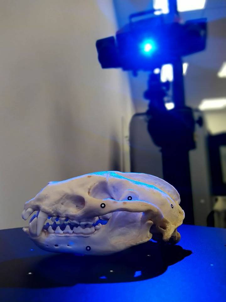
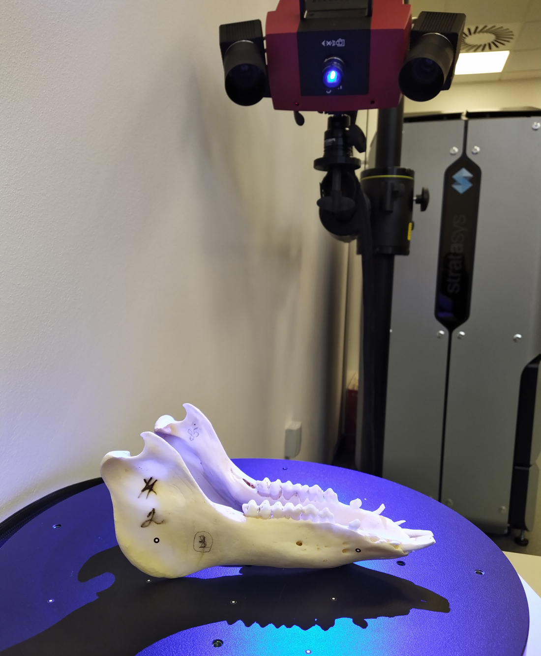
The second device is 3D scanner ATOS (Advanced Topometric Sensor) Compact Scan 12M Essential Line. This is a mobile contactless device consisting of stereo CCD cameras with a resolution of 12Mpx and a strip projector. This 3D scanner can scan an object with an accuracy of up to 0.01 mm, and the ATOS measuring system is certified for metrology. The device is placed on an adjustable stand and it has two measuring volumes - 300 and 600 mm. It is equipped with a blue light, enabling scanning regardless of the lighting conditions. The scanner uses the Fringe Projection method, in which precise stripes are projected onto the surface using a laser, deforming based on the shape of the object. The ATOS system uses a triangulation procedure. To achieve total digitization, the subject must be scanned from different angles. The ATOS system itself converts these captured images into a common coordinate system. The main advantage of the 3D scanner is its high resolution when scanning the surface of the object.
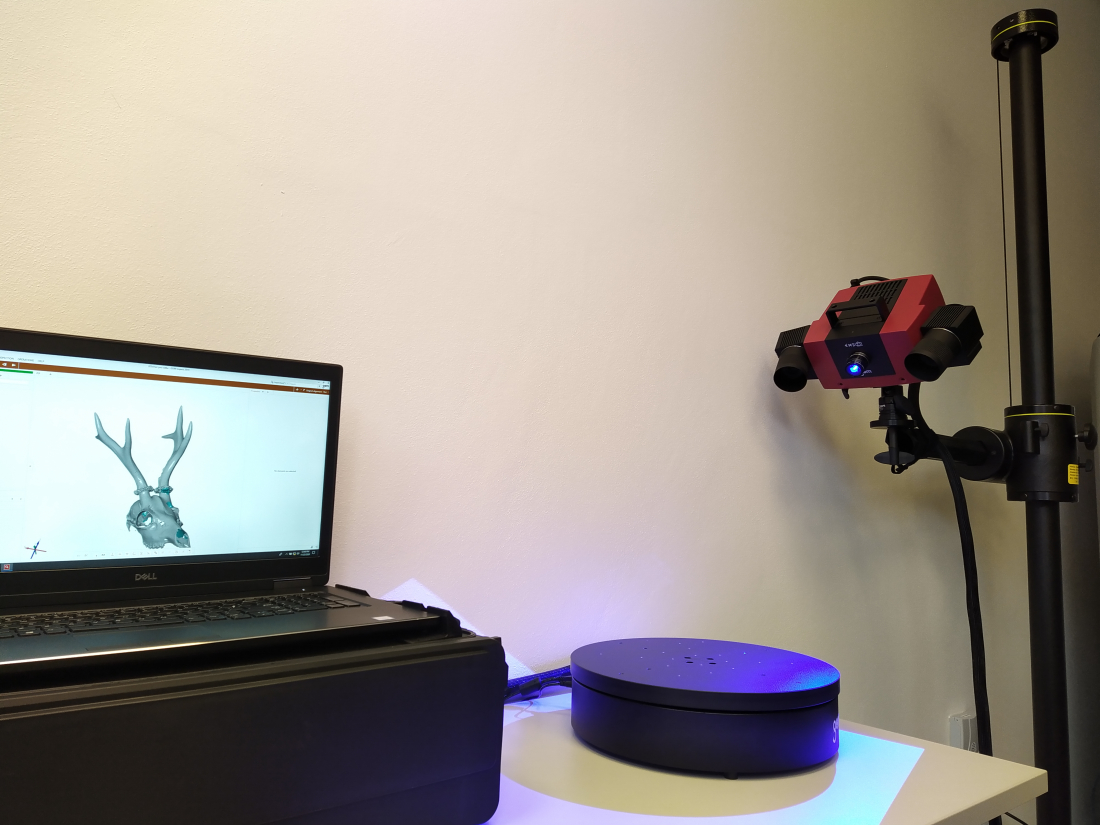
It is the combination of outputs obtained from both devices that provides unique results.
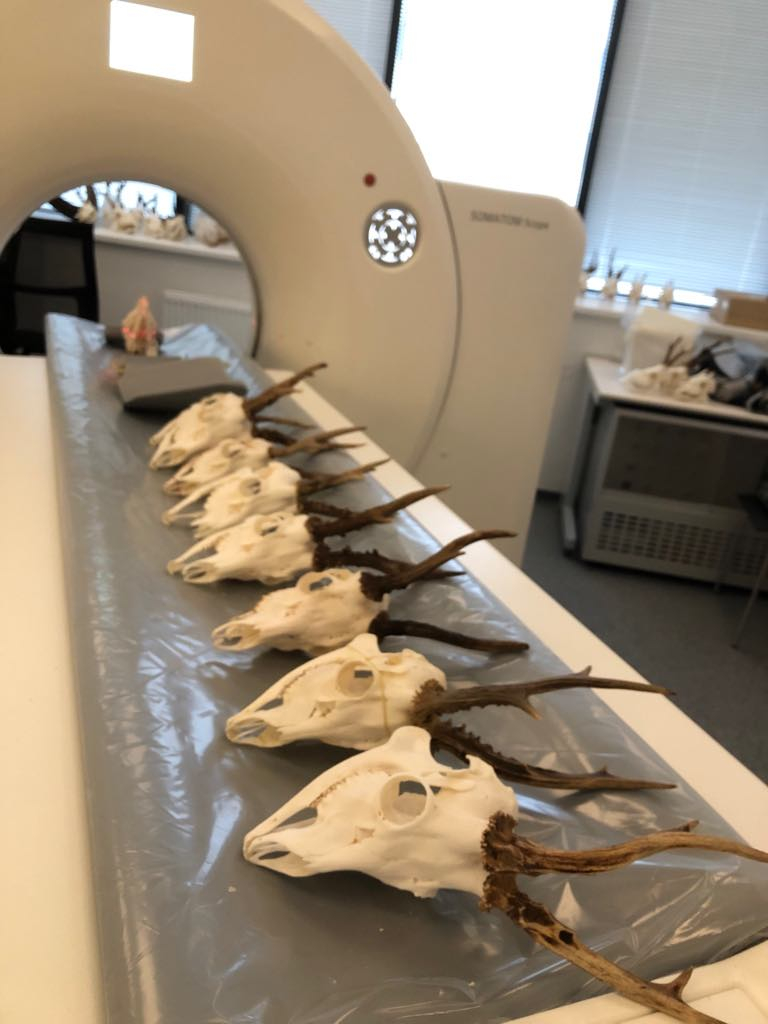
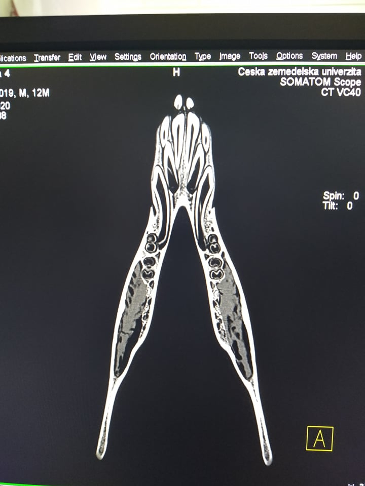
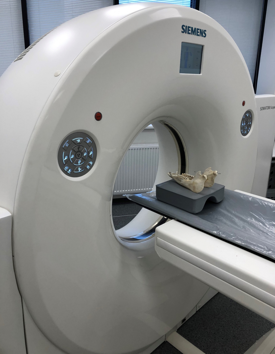
At present, we primarily focus on craniometry, odontology and the anatomy of game. The species of most interest are roe deer and wild boar, but we also focus on many others. The most common goal of craniometry is identification of the animal species by measuring the distances and dimensions between specified points on the vertebrate skull and determining its ethological or ecological connections. The basic technology used in craniometry is the measurement of dimensions using a caliper. Craniometric methods also include the measurement of antlers; basic quantities include length and size, but for some species, especially cervids (Cervidae), we also measure the volume of antlers. The volume of antlers is determined on the basis of methods recognized by the CIC (International Council for Game and Wildlife Conservation).