×
Department of game management and wildlife biology
banner

Laboratory work

Science, research and teaching > Laboratory work

   At the Department of Game Management and Wildlife Biology we have three new laboratory facilities: a brand-new biophysical behavioral lab with two magnetic coils, signal amplifiers, and recording equipment was created for conducting magnetoreception experiments in a highly controlled environment. Here we can set up all the important variables including light colour and intensity, strength, direction and density of magnetic fields, air and water temperature, etc.

   In each of two test rooms, animals are subjected to an artificial regulated magnetic field created by a coil beneath the box, or arena, in which they can move freely. The table beneath the arena also contains room for the installation of bar magnets. A high-resolution video camera is connected to the computer with analysing software and a powerful databank capable of storing terabytes of data. The walls and doors of the laboratory and test rooms are fully shielded against noise and strong magnetic fields produced by electronic equipment in the building. But that’s not all - the special material used for insulation also blocks radio waves from mobile phones, Wi-Fi, etc., which are known to counteract with certain types of magnetoreceptive processes. The animals in the test rooms are fully isolated from both the experimenter and the outer environment, allowing for complete control of experimental conditions crucial for extra sensitive magnetoreceptive reactions.

   The control room contains a sophisticated system of signal amplifiers and two powerful computers, where one or two experimenters can monitor the course of the test without affecting and disturbing the tested subjects.

   An example of experiments that can be conducted in the magnetic coil is a test of magnetic alignment. It is known that in calm conditions animals tend to spontaneously (uncontrollably and without additional stimuli) align their body along the N-S axis of a magnetic field. We can place an animal in the arena inside the coil, set the required parameters (strength, direction of the magnetic field, etc.), and then record the precise position of the animal’s body and its precise angle relative to the magnetic field’s axis.

   The animals used in the experiments should be safely and comfortably accommodated in between experiments. For this purpose, there is an animal lab in the neighbouring section. It consists of the aquarial room for keeping fish and amphibians, and a room with cages and plastic boxes for keeping rabbits, guinea pigs and small rodents. All parts of the equipment consist of non-magnetic material, or material shielded against magnetic fields. The walls of the animal lab are also magnetically shielded to remove all sources of interference with the controlled animal behaviour. Life support systems are at least partly controlled electronically.

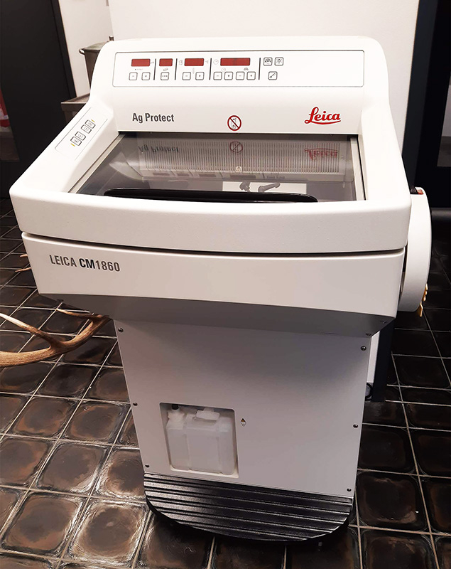
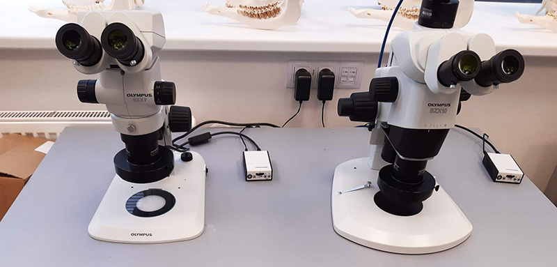
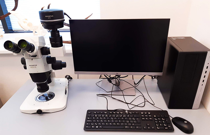
   Laboratory research involving high-resolution microscopy, molecular analyses and histological procedures is a brand-new trend in the activities of the Department of Game Management and Wildlife Biology. Science in the 21st century is extensively moving from macro- to micro-levels with the rapid development of methods of genome sequencing and protein analysis, as well as sophisticated computer technologies of bioimaging and image processing – and we as wildlife biologists need to keep up the pace. In collaboration with other departments and faculties of CULS, we established a new laboratory space for molecular research in the HiTech building and the FLD basement, where we have all means necessary to collect laboratory samples and preprocess them.

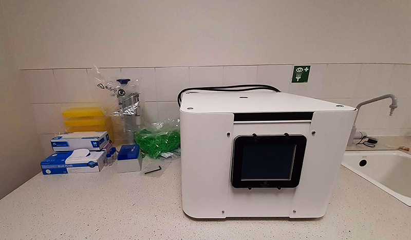
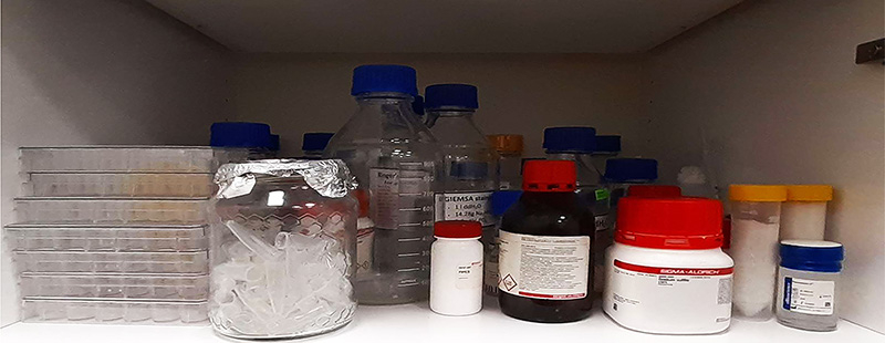
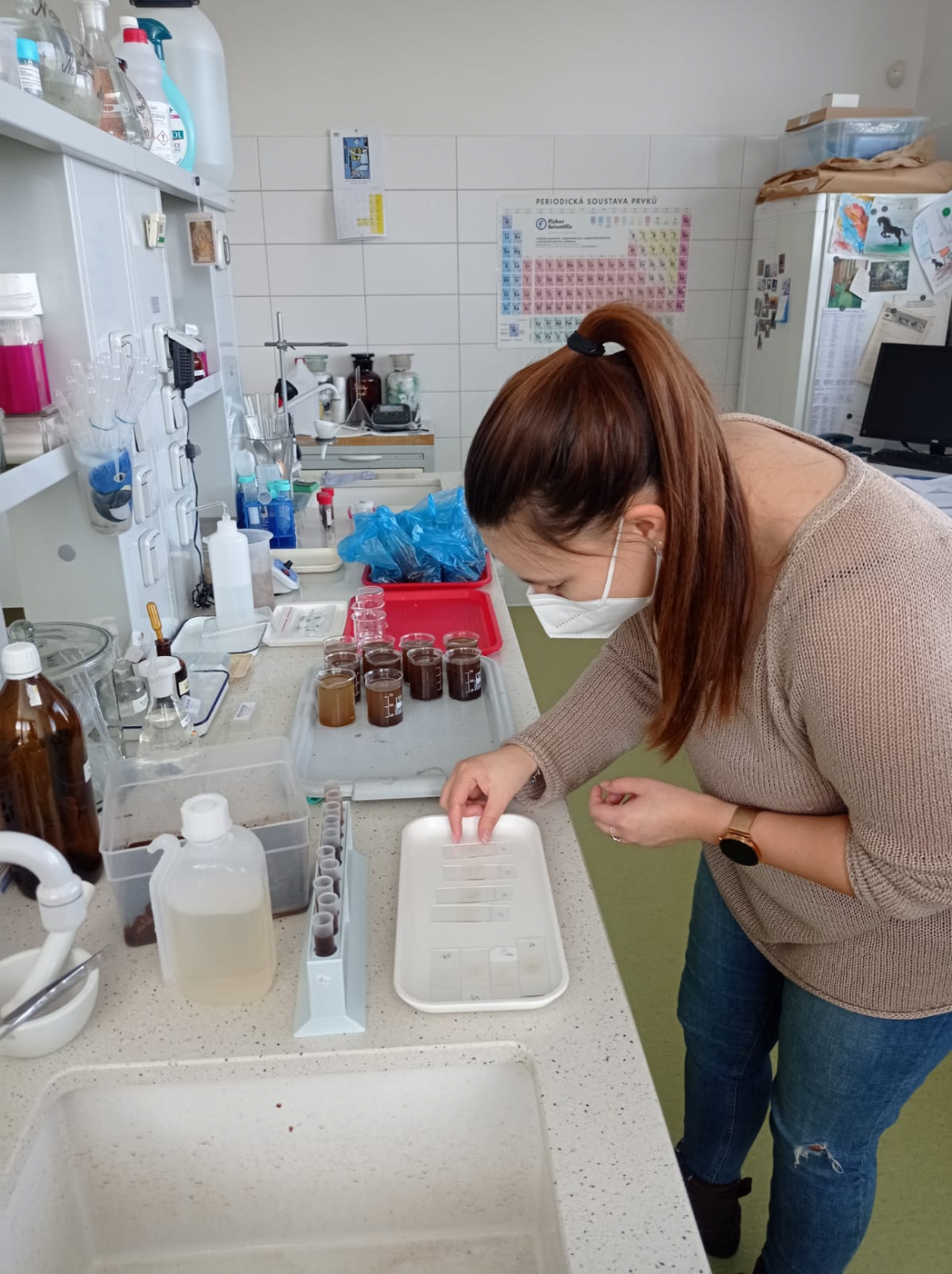
   In the lab next to the one with the coils and animal containment, we have a space for sample preparation, initial processing, and storage. Samples of blood, feaces, urine, and biological tissues that are collected to monitor the physiological state of the animals in the experiment are stored frozen in voluminous powerful -25°C freezers. Next to them are laboratory tables with equipment for homogenizing, centrifugation and pipetting, allowing the extraction of substances of interest (DNA, RNA, proteins, steroid hormones, etc.) from raw samples for their further processing in specialized molecular labs in CZU or other institutions.

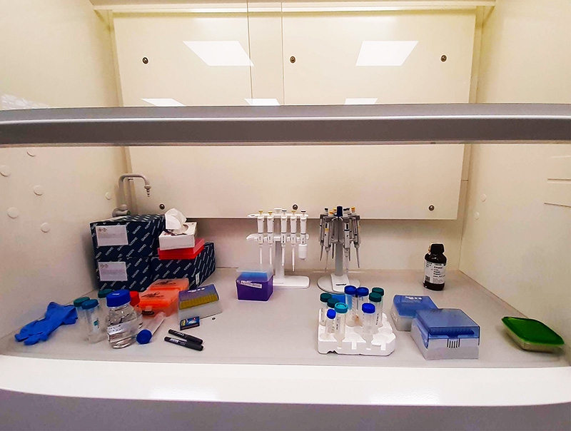
   Our lab therefore allows the preparation of samples for DNA sequencing used for various purposes from species identification and barcoding to complex phylogeographical analyses. We can also safely preserve tissues for RNA extraction and sequencing to study the expression of specific genes linked to behavioural reactions, or extract steroid hormones (cortisol, DHEA etc.) for stress experiments performed in magnetic coils or in nature.

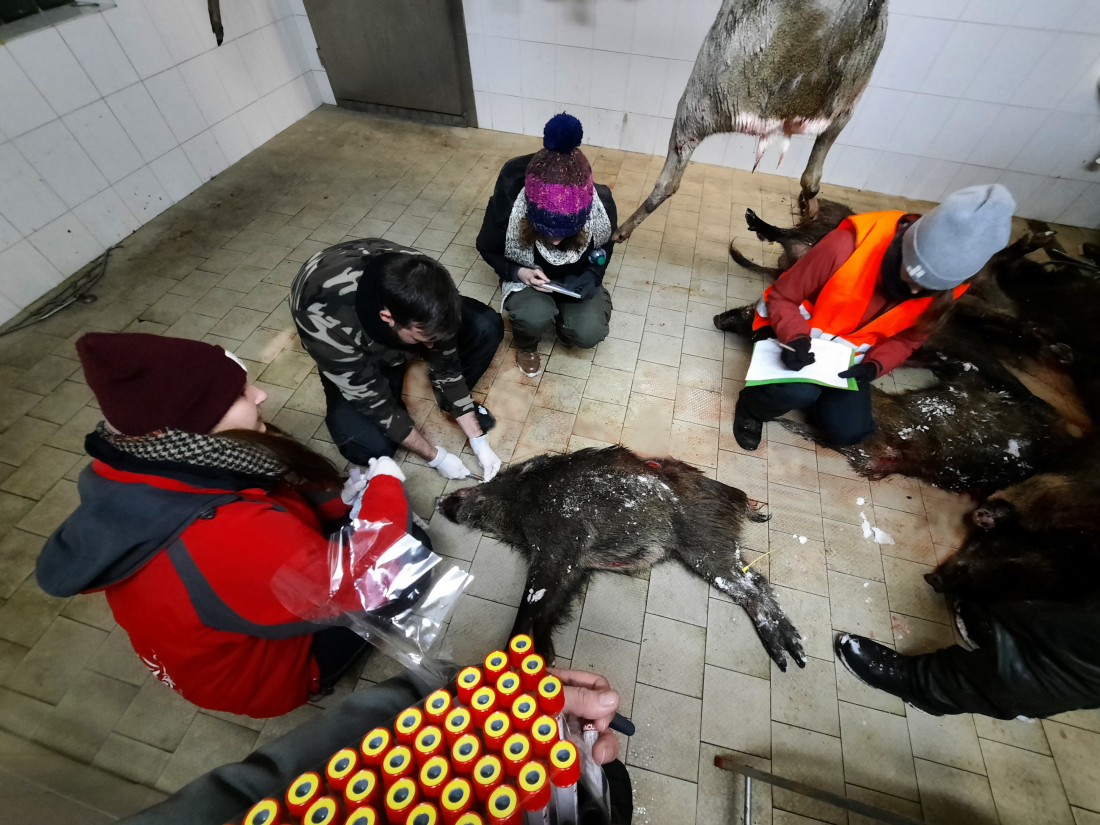
   For example, to measure steroid hormone levels in wild boar, we collect blood samples that should first be centrifugated to separate the serum with steroids from blood cells. We also collect feaces that are dried in speedvac, homogenized and weighted before the extraction. The process of extraction is relatively uniform for different types of samples in terms of chemicals used (ethylacetate or pure ethanol). The specific protocols consist of multiple sequences of pipetting, vortexing, centrifugation and speedvac drying in a variety of combinations. The extracted steroid pellets are then deep-frozen until used for ELISA.

   For DNA and RNA extraction from fresh or ethanol-fixed samples, we use commercial kits that allow us to thoroughly separate nucleic acids from fatty or protein compounds, clean and concentrated samples to assure high quality amplification.

   In the Hi-Tech building, we have brand-new equipment for light microscopy and animal histology. Certain types of animal tissue, particularly brain tissue, require very gentle preparation of slices with a minimum of fixation procedures. For this purpose, we have a new Leica CM1860 cryotome (freezing microtome) that allows us to prepare slices in a thickness up to 1 µm. Another modern device for delicate or immunohistochemically stained tissues, vibratome (vibration microtome for cutting agarose-embedded samples) will be added to our equipment very soon.

   To obtain high-quality histological images, carefully prepared tissue samples and slides are required. In our lab, we have new high-quality histological equipment for staining and drying biological material, and a vast selection of top-quality chemicals from leading companies (Merc, Invitrogen, Sigma Aldrich etc.) for conventional and fluorescent histochemistry.

   A set of high-quality OLYMPUS light microscopes with built-in digital color photo/videocameras allows us to obtain high-quality tissue images fixed and stained by conventional methods. We also plan to extend the spectrum of histology methods to various immunofluorescent staining techniques, allowing detection of the smallest and most eluding organismal structures that may be used for complex and sensitive reactions such as magnetoreception.

Samples from hunting events

   Stress is an important factor in animal welfare. Longstanding stress research focuses on humans and livestock, whereas stress in wildlife, and especially game species, is equally important. Both natural and human-induced stress triggers, such as weather changes and tourism, are increasing. Urban sprawl is promoting human-wildlife conflict, which results in behavioural and physical changes, including a stress response.

   Assessing animal welfare and disease surveillance in free ranging wildlife species can be quite challenging. Evaluating stress essentially requires blood or feaces from dead or captured animals. For most analyses, samples need to be fresh and uncontaminated. Therefore, in free ranging game species, sampling is the easiest after hunting events. Especially driven hunts enable easy sampling in large quantities. In the Department of Game Management and Wildlife Biology, we sample blood from shot wild boar by puncturing the cavernous sinus. This facilitates quick and clean blood samples in a sufficient volume for various analyses. The samples are primarily used to measure hormone levels, as well as other parameters, such as haematocrit and glucose levels. The data are used to evaluate the stress levels of individuals during hunting events, as well as long term stress. Combining different parameters gives a gross assessment of the current health condition of an animal, and it also enables some reconstruction of past conditions to check for time-, situation- and location-dependent differences.

Parasite infestation

   Population densities and behaviour of game animals often fluctuates due to the transmission of parasites and diseases. Parasites are organisms that live in or on a host, feeding from or at the expense of its host and using it for reproduction. It negatively affects the well-being of the host, and it can result in behavioural changes. The detection of parasites is also important for meat quality assessment. For this reason, the tissue of hunted animals is inspected for visible signs of parasites, and blood and tissue can be examined in laboratories. Parasitic worms (helminthes) are one of the most common parasites. Adults can be seen with the naked eye, making it easy to detect them when eviscerating hunted game. Given the fact that most helminths infect the gastrointestinal tract, eggs can be found in the host’s feaces.

   The Department of Game Management and Wildlife Biology uses this to non-invasively detect and monitor parasite infestation and transmission in free ranging wild boar groups in selected areas in the Czech Republic. Groups can be identified using GPS collared individuals known to move in groups. The helminth parasite load is estimated using semiquantitative counting methods. Using simple flotation and sedimentation methods, we were able to detect eggs of helminth species such as Ascaris suum (Fig. 1), Capillaria sp. (Fig. 2), Metastrongylus sp. (Fig. 3 top) and Strongyles (Fig. 3 bottom), which are common wild boar parasites.